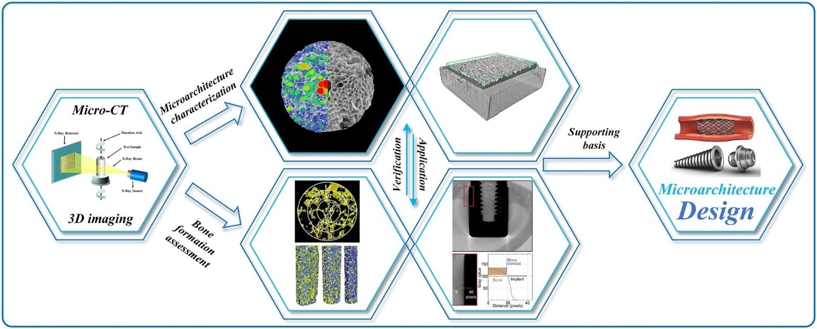
As an advanced microscopy technology with strong sample adaptability and non-destructive three-dimensional (3D) characteristics, X-ray micro-computed tomography (Micro-CT) can establish the overall connection between various microarchitecture parameters and accelerate the research process of porous metallic implants and scaffolds.
In this review, the Micro-CT based quantitative evaluation methods of microarchitecture and bone formation are investigated. To ensure reliability of the results, the Micro-CT setup is discussed briefly and the essential image processing algorithms are introduced in detail. The significance and limitations of Micro-CT are analyzed in the context of research on porous metallic implants. We also discuss the future development of Micro-CT technology in the field of biological tissue engineering.