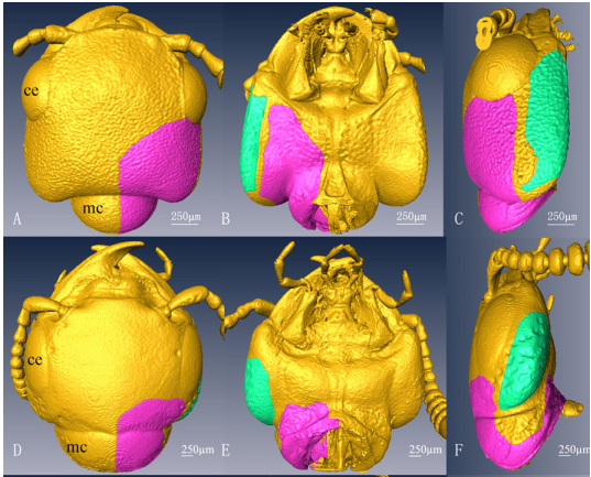
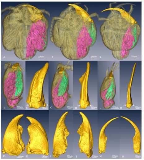
X-ray micro-CT is a powerful tool to visualize without damage details of the inner structures of beetles, the largest order of insects with a hard external skeleton. This contribution shows the three-dimensional (3D) reconstruction of the head morphology of three rove beetle species (Insecta, Coleoptera, Staphylinidae)—Noddia sp., Creophilus maxillosus, and Hesperosoma sp.—using X-ray microtomography at a spatial resolution of 6 μm. The details of skeletal muscle fiber insertions are described, giving a comprehensive overview of mandible mobility and organization. With the support of 3D rendering, we discuss the relationship among the mandible forms, the development of the muscles controlling the movement, and the head morphology. The well-developed posterior part of the head capsule is always accompanied by a well-developed mandible, a large adductor muscle, and a large apodeme for the wide areas of the muscle fiber attachment. In Noddia sp., muscles connected to the posterolateral angle of the head capsule are mainly short muscles, whereas in Creophilus maxillosus, the latter are mainly long muscles, and in Hesperosoma sp. no mandible adductor muscle fibers are present on the posterolateral angle of the head capsule. These results offer new invaluable information regarding the biting functions of beetle mandibles and the trend of their morphological change during their long-term evolution.
|
Fig. 2 Three dimensional reconstructions of Noddia sp. (A–E), Creophilus maxillosus (F–J), and Hesperosoma sp. (K–O). A, B, F, G, K, L Head, transparent view; mandible and mandible muscles, shade view; A, F, K dorsal view; B, G, L lateral view. C–E, H–J, M–O Mandible, shade view; C, H, M lateral view; D, I, N ventral view; E, J, O dorsal view. Purple the mandible closer muscle, green the mandible opener muscle. Arrows show mandible insertions with the head capsule. Between arrows we show the mandible rotation axis |