
Abstract: One of the challenges of the study of geological samples is obtaining volumetric mineralogical properties of the rocks. As a part of special core analysis, total elemental (mineralogical) content of a rock can be measured on the crushed rock using classical X-ray diffraction and X-ray florescence techniques. A new generation of advance core analysis techniques such as specialized scanning electron microscopy and Raman scanning microscopy can be used to obtain two-dimensional elemental composition of the rock [0, 00] which can be propagated into three dimensions, but these are still not genuine experimental techniques for non-destructive 3D chemical testing of the material. In this work we have investigated the applicability of a combined X-ray micro-computed tomography (micro-CT) and micro X-ray fluorescence (micro-XRF) system for obtaining information on the spatial distribution of chemical elements in a rock sample. The unique capabilities of the technique are discussed together with its limitations.
Keywords: micro CT / X-ray / fluorescence / mineralogical / electron microscopy / geological / rock
For full text click here .
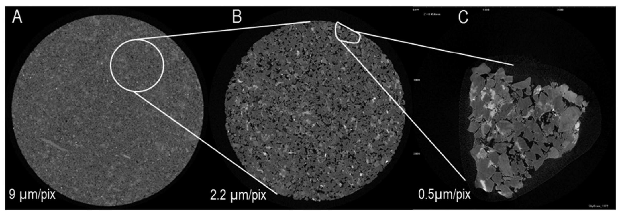
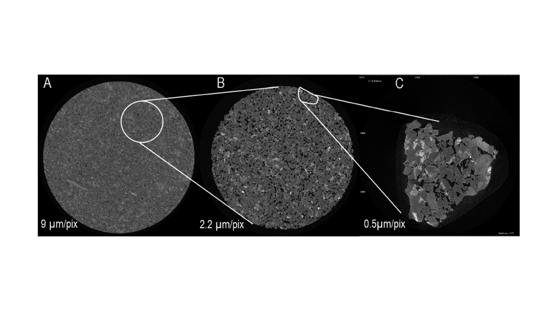
X-ray micro-computed tomography data from a sandstone core plug: XY slices of the sample scanned at different resolutions are shown. (A) 30 mm diameter plug scanned at a resolution of 9 μm/pixel. (B) 8 mm diameter plug at 22 μm/pixel. (C) 2 mm chip of the rock at 0.5 μm/pixel. All data were obtained using a standard Al+Cu filter
.png)