Abstract
Introduction: Odontoid fractures easily lead to instability, causing spinal cord injury. The aim of this study was to measure and analyze the micro-architecture and morphometric parameters of the normal odontoid with high-resolution three-dimensional (3D) micro-computed tomography (micro-CT).
Methods: Micro-CT scans were obtained from five normal odontoid processes. The scanned data were reconstructed with micro-CT software, and the nutrient foramina and the ossification center of the base of the odontoid were revealed. The trabeculae of the odontoid were measured and divided into four parts to obtain the volume fraction of regions of interest.
Results: High-resolution 3D images of the micro-structures' parameters were obtained from the odontoid using micro-CT software. The images demonstrated sponge-like trabecular bone, with the trabeculae showing a complex, net-like micro-construction. The subchondral bone plate was of lamella-like, compact construction and extended and transformed into a net-like structure with rod-shaped trabeculae arranged radially in all directions. There was a statistically significant difference in the volume fraction compared with the region of interest in the fourth part of the trabeculae and the first part of the odontoid (P < 0.05). The nutrient foramina and the ossification center of the odontoid were also observed.
Conclusions: It is feasible to use high-resolution 3D micro-CT to evaluate the micro-architecture of the normal odontoid. Other studies can benefit from use of the micro-CT images, such as finite element evaluations.
Keywords: Axis, micro-architecture, micro-computed tomography, odontoid.

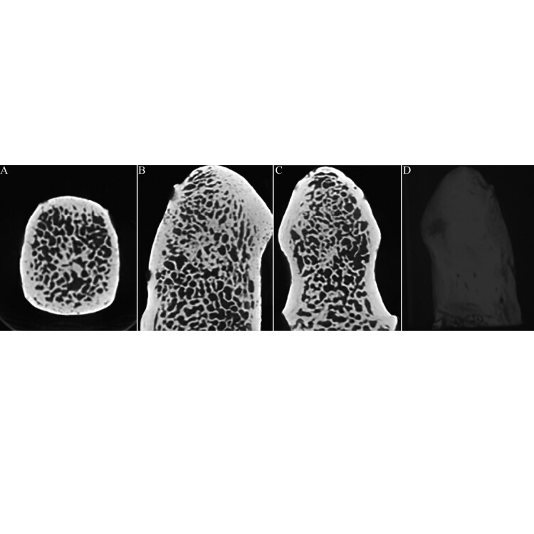
Figure 1 Micro-computed tomography (CT) images of the odontoid. (A: cross section, B: sagittal section, C: coronal section, D: three-dimensional, 3D).

Figure 3 (A) Type II fracture; (B) 3D-Micro-computed tomography shows that low bone volume fractions represent weak points in the structure of part IV.